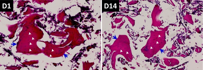
Figure 6. HE staining and evaluation of co-culture of BMSCs with composite scaffold constructed using Si-CaP, autogenous fine particulate bone powder, and alginate.
Left panel: 1-day after co-culture; Right panel: 14-days after co-culture. These images, especially at 14-days, autogenous fine particulate bone powder and Si-CaP are distributed uniformly, and the bone lacuna (blue arrow heads) and osteocytes within the bone matrix are clearly obseved with an integral structure, the nucleus of osteocytes (white arrow heads) are deep blue stained, indicating osteocytes live well under current experimental condition.

