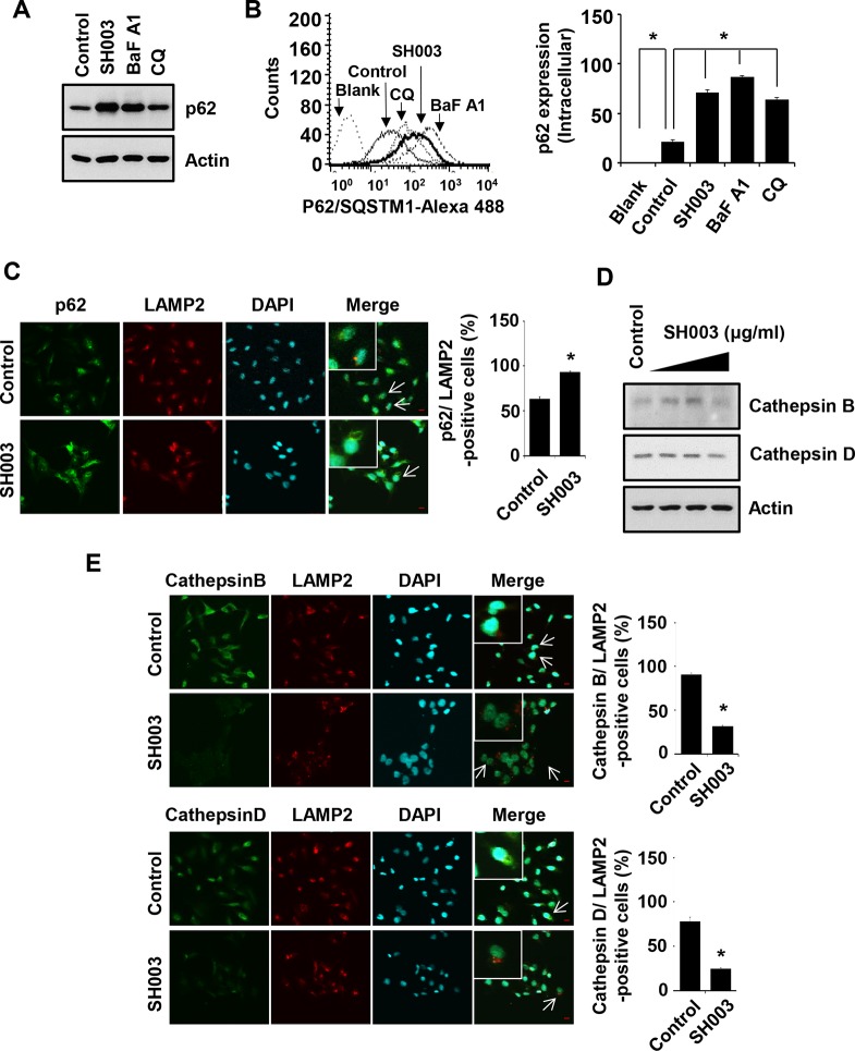
Figure 5. SH003 induces p62 accumulation via reduction of Cathepsin expression.
(A) MDA-MB-231 cells were treated with SH003 (500 μg/ml), BaFA1 (400 nM) and CQ (10 μM) for 24 hours. p62 protein expression was objected with western blots. Actin was used for the internal control. (B) Cells were treated with SH003 and autophagy inhibitors (BaFA1 and CQ) for 24 hours and stained with p62 -Alexa 488-conjugated p62 antibody for 30 minutes. p62 accumulation in the cells were detected using FACSCalibur. *P < 0.05. (C) MDA-MB-231 cells were treated with SH003 for 24 hours and stained with p62 (1 μg/ml) and LAMP2. DAPI was used as nucleus staining. The object was 20× and scale bar indicates 10 μm. *P < 0.05. (D) Cells were treated with SH003 for 24 hours and then performed western blots with anti-Cathepsin B and -Cathepsin D. Actin was used for loading control. (E) MDA-MB-231 cells were treated with 500 μg/ml of SH003 for 24 hours and stained with Cathepsin B (1:50)/LAMP2 and Cathepsin D (1:50)/LAMP2. Images were obtained with using Olympys FV10i Self Contained Confocal Laser System. The object was 20× and scale bar indicates 10 μm. *P < 0.05. Experiments were performed in triplicate. Bars indicate means that standard deviations (SD).

