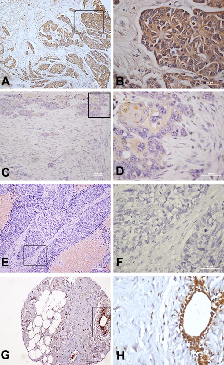
Figure 1. Immunohistochemical staining of TLR5 protein in breast cancer tissues.

(A-B) High expression of TLR5 in breast cancer tissue (10× 40×). (C-D) Low expression of TLR5 in breast cancer tissue (10× 40×). (E-F) Negative expression of TLR5 in breast cancer tissue (10× 40×). (G-H) TLR5 expression on tumor-adjacent breast tissue (10× 40×).
