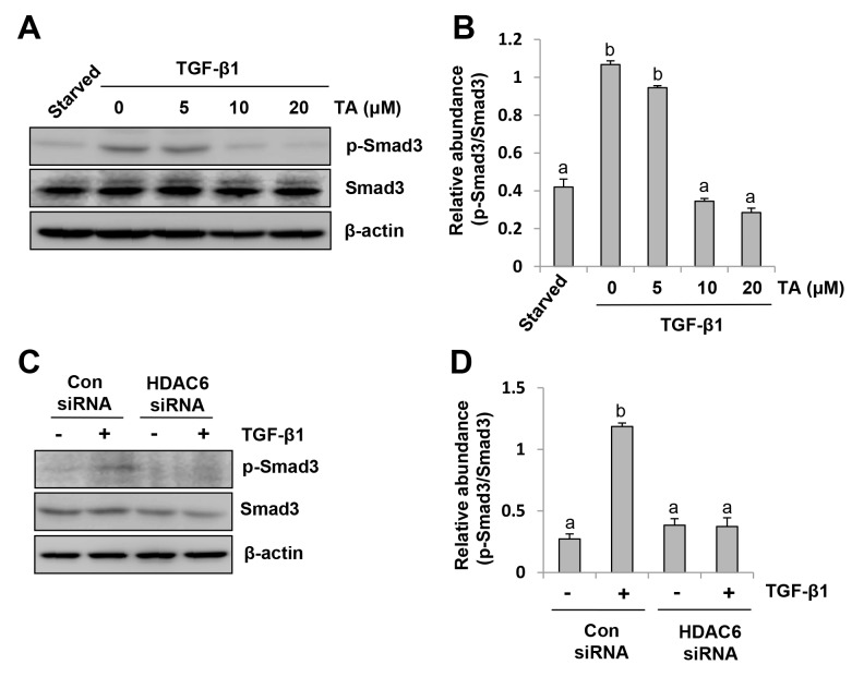
Figure 3. HDAC6 is required for TGF-β induced Smad3 phosphorylation in peritoneal mesothelial cells.
(A) Serum-starved HPMCs were pretreated with various concentrations of TA (0-20 μM) for 1 hour and then exposed to TGF-β1 (10 ng/ml) for an additional 24 hours. Cell lysates were subjected to immunoblot analysis with antibodies against p-Smad3, Smad3, or β-actin. (B) Expression level of p-Smad3 was quantified by densitometry and normalized with Smad3. (C) Serum-starved HPMCs were transferred with siRNA targeting HDAC6 or scrambled siRNA and then incubated with TGF-β1 (10 ng/ml) for an additional 24 hours. Cell lysates were subjected to immunoblot analysis with antibodies against p-Smad3, Smad3, or β-actin. (D) Expression level of p-Smad3 was quantified by densitometry and normalized with Smad3. Values are means±SD of at least three independent experiments. Bars with different letters (a-d) for each molecule are significantly different from one another (P<0.05).

