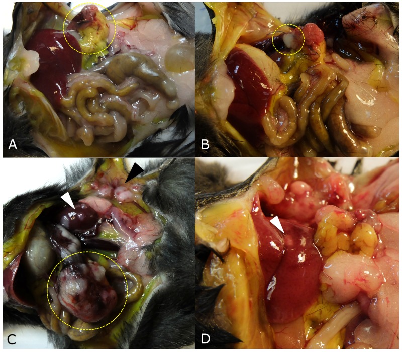
Figure 3. Tumor growth patterns at laparotomy.
(A) The implanted tumor disappeared and only peritoneal fat was attached around surgical bed in one mouse (yellow dotted circle). (B) If the tumor grew to less than 5 mm, the tumor growth pattern was defined as ‘no growth’ (dotted circle) in a mouse in the control group. (C) Large pancreatic tumor (yellow dotted circle), kidney metastasis (white arrow head), and peritoneal seeding (black arrow head) were observed in a mouse in the splenectomy group. (D) Liver metastasis was observed in one mouse (white arrow head).

