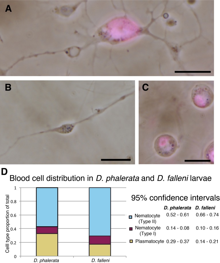
Fig 1. Three distinct hemocytes are found in 3rd instar larvae of Drosophila phalerata and Drosophila falleni.
Live cell imaging of three cell types, counter stained with DAPI in magenta, shows two classes of nematocytes. Nucleated nematocyte (type I) with strong DAPI signal and spindle projections extruding from the cell body (A), and enucleated nematocyte (type II) lacking DAPI staining but morphologically similar, having spindle projections extending from central cellular region (B). DAPI positive plasmatocytes are also found in circulating hemolymph (C). The distribution of hemocyte populations in each species was quantified (D), with type II nematocytes being the most abundant cell type in both species. Scale bars are 10 microns (A-C).

