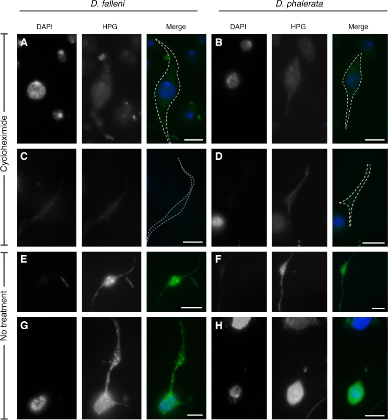
Fig 4. Hemocytes are able to synthesize protein.
Nascent protein synthesis was visualized using the Click-it protein synthesis kit, measured with green fluorescence (HPG). DNA is marked with DAPI staining. As expected, type-I nematocytes (A & B) and type-II nematocytes (C & D) in the cycloheximide treatment did not indicate significant levels of protein synthesis. However, these cells without cycloheximide treatment had green fluorescence, signifying protein synthesis. White dashed line in merged images denotes the cell outline as based on phase-contrast image. Images were taken at identical microscope and camera settings with standard fluorescent microscope. Scale bars are 10 microns.

