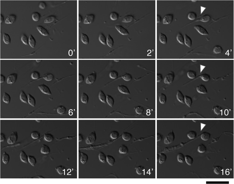
Fig 7. Live imaging captures formation of type-II nematocyte.
Time-lapse imaging shows the formation of a spindle-like projection and an eventual pinching off of this appendage from the progenitor cell (T12). This budded cell maintains dynamic movement after separation. Time intervals are 2 minutes with DIC microscopy and perfect focus. White carrot indicates specific region of interest. Scale bar is 20 microns.

