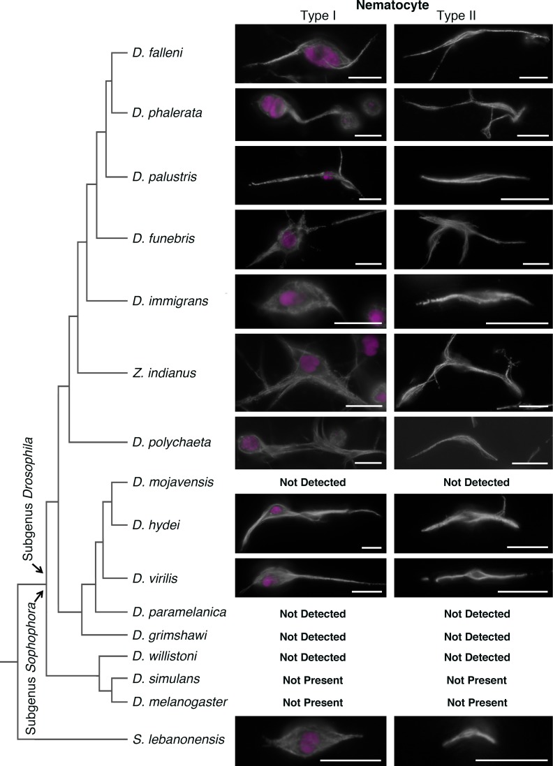
Fig 8. Numerous Drosophila species have type-I and type-II nematocytes.
Hemolymph from multiple Drosophila species was examined for the presences of type-I and type-II nematocytes. Images show tubulin (white) and DAPI (magenta). “Not Detected” reports that the cell type was not detected from multiples samples in this study. In D. melanogaster and D. simulans these cell types are assigned a “Not Present” status, reflecting that these cell types are not present based both on the current findings and supported by a significant body of literature. The left panel reports a phylogenetic tree for these species; however, horizontal length of the branches does not represent a quantified genetic distance or evolutionary time. Scale bars are 10 microns.

