
Figure 1. DAI is essential for IAV-induced cell death in MEFs and alveolar epithelial cells.
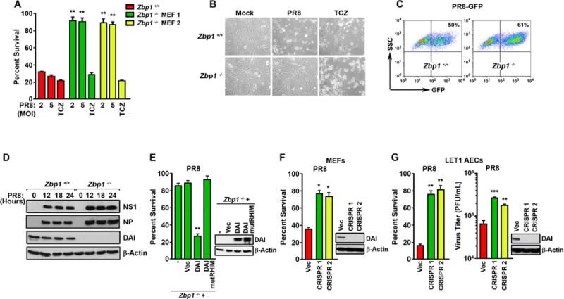
(A) Zbp1+/+ or zbp1−/− MEFs from two separately-maintained colonies (MEF 1 and MEF 2) were infected with PR8 (MOI=2 and 5), or treated with the combination of TNF-α [50ng/ml] + cycloheximide [250ng/ml] + zVAD [50uM] (TCZ) and cell viability was determined 24 hr p.i. (B) Photomicrographs of zbp1+/+ and zbp1−/− MEFs infected with PR8 (MOI=2) or treated with TCZ for 24 hr. (C) FACS analysis of zbp1+/+ and zbp1−/− MEFs infected with PR8-GFP (MOI=2) for 18 hr. The y-axis shows side scatter. Mock-infected cells showed negligible (<0.1%) GFP-positivity. (D) Lysates from zbp1+/+ and zbp1−/− MEFs infected with PR8 (MOI=2) were examined for expression of NS1, NP, and DAI. (E) Immortalized zbp1−/− MEFs reconstituted with empty vector (Vec), full-length murine DAI (DAI), or murine DAI with mutant RHIM domain (amino acids 192–195 IQIG to AAAA) (DAI mutRHIM) were infected with PR8 (MOI=2) and cell viability was determined 24 hr p.i. Expression of WT or mutant DAI in these cells is shown to the right. (F) WT MEFs in which DAI expression was ablated by CRISPR/Cas9 targeting were infected with PR8 (MOI=2) and cell viability was determined 24 hr p.i. Ablation of DAI expression was confirmed by immunoblotting (right). (G) LET1 cells in which DAI expression was ablated by CRISPR/Cas9 targeting were infected with PR8 (MOI=2) and cell viability was determined 12 hr p.i. (left). In parallel, progeny virion output from these cells was determined 30 hr p.i. (right). Viability data shown in this figure are representative of at least three independent experiments. Error bars represent mean +/− SD. *p<0.05, **p<0.005, ***p<0.0005. See also Fig. S1.