
Figure 2. Faster oscillations of p53 in mouse cells are not due to the p53 sequence, general metabolic state or DNA damage signaling.
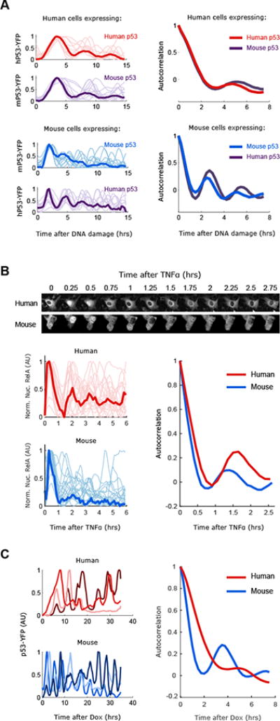
(A) Human (MCF7) or Mouse (NIH3T3) cells expressing either human or mouse p53-YFP were DNA damaged (NCS 100ng/ml) and imaged for 15hrs (faint lines indicate single cells, bold lines indicate the average). Oscillations of p53 were quantified and the period was calculated with autocorrelation function (N>20 cells). (B) Human (MCF7) or Mouse (NIH3T3) cells expressing RELA-CFP were treated with TNFα (10ng/ml) and imaged for 6hrs. RelA-CFP nuclear signal was quantified and autocorrelations was used to calculate the oscillatory period (N>20 cells). (C) Human (MCF7) or Mouse (HEPA1c1c7) cells expressing a dox inducible p53-YFP construct were imaged after addition of doxycycline (50ng/ml). Three examples traces are shown and the quantified periods (N>10 cells).