
Figure 1.
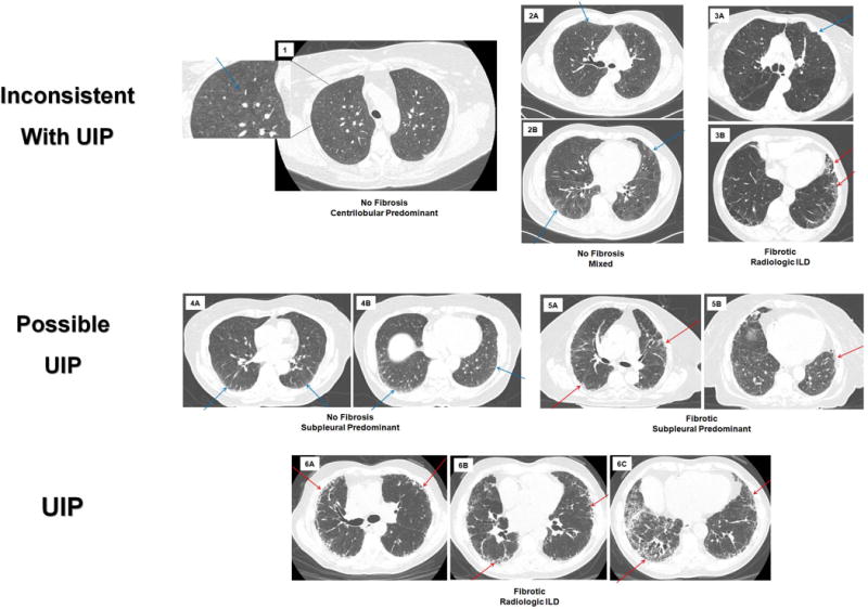
Chest computed tomographic (CT) images depicting radiologic subtypes and the overlap between subtypes of interstitial lung abnormalities (ILA). In all panels the blue arrows point to areas of ILA without fibrosis, the red arrows point to areas of ILA with fibrosis, each panel (1–6) represents one participant. Panels 1–3 demonstrate patterns of ILA that are inconsistent with usual interstitial pneumonia (UIP), panels 4–5 demonstrate patterns of ILA that are possible UIP and panel 6 is a pattern of ILA that is consistent with UIP. Panel 1 represents non-fibrotic, centrilobular predominant ILA, with an area zoomed in to highlight the centrilobular ground glass nodules. Panel 2 is non-fibrotic, mixed pattern of ILA, in 2A the blue arrow points to subpleural reticulation; in 2B the arrows demonstrate both subpleural and centrilobular ground glass. Panel 3 is fibrotic (see the red arrows in 3B), radiologic interstitial lung disease (ILD), that is inconsistent with UIP due to the pleural plaque (blue arrow) in 3A. Panel 4 is non-fibrotic, subpleural predominant ILA, blue arrows pointing to subpleural reticulation. Panel 5 is fibrotic, subpleural predominant ILA, with red arrows in both panels pointing to traction bronchiectasis. Panel 6 is fibrotic, radiologic ILD; red arrows highlight traction bronchiectasis and honeycombing.