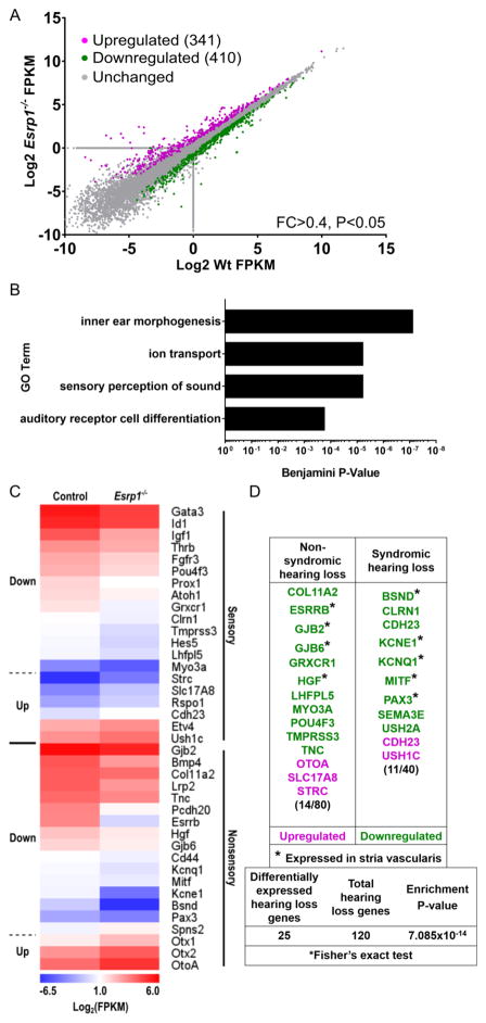
Figure 3. Sensory and nonsensory gene expression profiles are disrupted in the cochlear epithelium of Esrp1−/− embryos.
(A) Plot of differentially expressed genes between wild type and Esrp1−/− cochlear epithelium at E16.5 with a fold change (FC) > 0.4 (n=3 replicates, P<0.05). (B) Gene Ontology term enrichment for differentially expressed genes between wild type and Esrp1−/− cochlear epithelium. (C) Heatmap of differentially expressed genes separated into sensory and nonsensory categories. (D) Hearing loss genes are significantly enriched in the set of differentially expressed transcripts between control and Esrp1−/− mutants. Genes expressed in the stria vascularis are marked with an asterisk.

