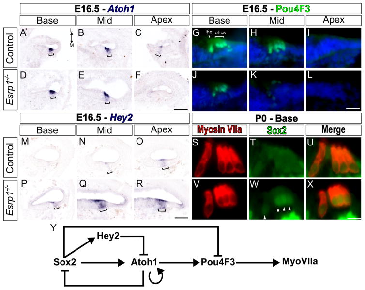
Figure 4. Hair cell differentiation is delayed in Esrp1−/− embryos.
(A–X) Transverse sections through defined regions of the cochlear duct (Base, Mid, Apex) from control and Esrp1−/− embryos stained for Atoh1 mRNA (A–F), Pou4F3 protein (G–L) and Hey2 mRNA (M–R) at E16.5 (n=5 or 6). Staining in prosensory domain is marked with a bracket. Weak Atoh1 expression at the apex of the cochlear duct in control embryos (C) is consistently absent in Esrp1−/− embryos at this stage. (S–X) Transverse sections through the organ of Corti of control and Esrp1−/− newborn pups (P0) stained for MyoVIIa and Sox2. Sox2 staining persists in hair cell nuclei of Esrp1−/− embryos (arrow heads in W). Scale bar = 100μm (A–F, M–R), 10μm (G–L) and 5μm (S–X). (Y) Schematic of the gene regulatory network controlling hair cell differentiation. Abbreviations: inner hair cell (ihc), outer hair cells (ohc), medial (M), lateral (L). See also Figures S4 and S5 and Table S2.

