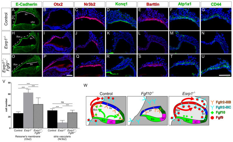
Figure 7. Ectopic signaling through Fgf9/Fgfr2-IIIc is responsible for the lateral cochlear wall defects in Esrp1 mutants.
(A–U) Transverse sections through the cochlear duct of control (n=15), Esrp1−/− (n=7) and Esrp1−/−;Fgf9+/− (n=6) embryos at E18.5 immunostained for E-cadherin (A,H,O), Otx2 (B,I,P) and cell type specific markers of the stria vascularis (C–G, J–N, Q–U). Scale bars = 50μm. (V) Quantification of cells expressing Otx2 and Nr3b2 represented as mean ± SD (***P<0.0001, ANOVA with Tukey’s test). (W) Schematic representation of the lateral cochlear wall phenotypes manifesting from altered Fgf signaling in Fgf10−/− and Esrp1−/− mutants compared to a control embryo. See also Figure S6.

