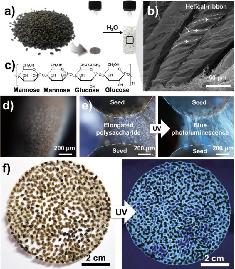
Fig. 1.
a) An optical image of basil seeds and their water-swelling behavior. b) Scanning electron image of the hydrocolloid of basil seed. c) Chemical structure of the representative polysaccharide in basil seed (i.e., glucomannan) d) Fluorescence images of the outer layer of an untreated basil seed. e) Fluorescence microscope images and f) optical images of a swelled basil seed cluster (λex = 312 nm).

