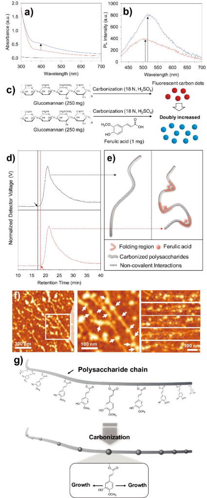
Fig. 4.
a) UV-Vis absorption of natural glucomannan before carbonization (black), after carbonization (red), and the addition of ferulic acid to glucomannan followed by carbonization (blue). b) Photoluminescence (ëex = 400 nm) measurement of the carbonized glucomannan in the presence (blue) or absence (red) of ferulic acid. c) The schematic experimental procedures and the illustration in which carbon dots are doubly increased in presence of a trace amount of ferulic acid during carbonization, based on the spectroscopic results from UV-vis and photoluminescence. d) The GPC results of carbonized natural glucomannan in the absence (black) or presence of ferulic acid (red) and e) the corresponding schematic illustration to explain the role of ferulic acid. f) The representative AFM image with corresponding magnified image, and additional images of the carbonized polysaccharides from basil seed. Developed carbon dots from polysaccharides were marked as arrows and circles. g) Proposed seed-growth mechanism of carbon dots from natural polysaccharides.

