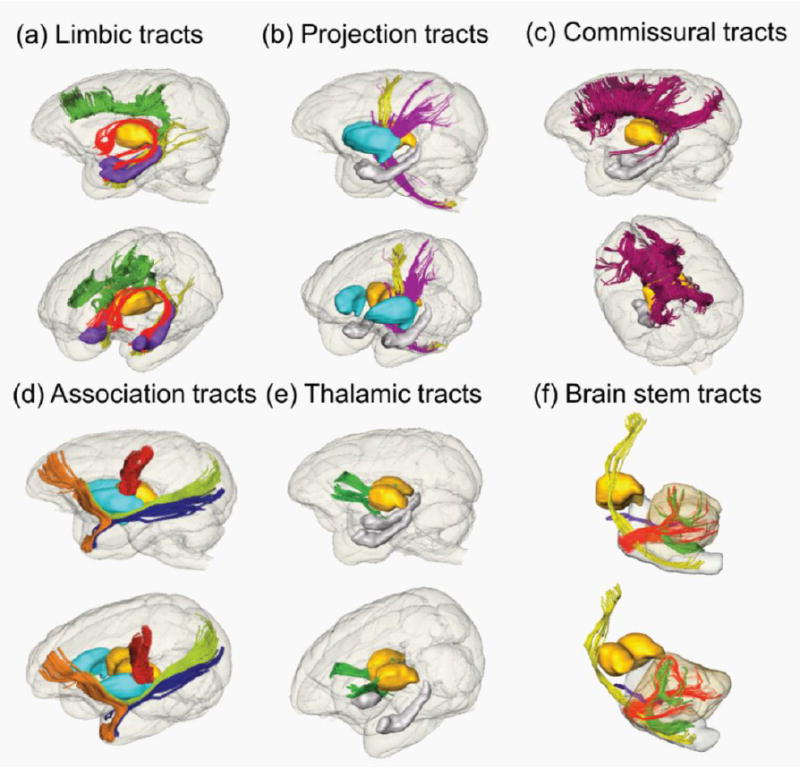
Figure 3.

3D reconstructed limbic (a), projection (b), commissural (c), association (d), thalamic (e) and brain stem (f) tracts of a macaque brain. Lateral and oblique views are displayed for each panel. Reconstructed tracts are cingulum in the cingulate gyrus (green), cingulum to the hippocampus (yellow) and fornix (red) in (a); corticospinal tract (yellow) and cerebral peduncle (purple) in (b); corpus callosum including genu, body, splenium and tapetum projecting to the temporal lobe (crimson) in (c); uncinate fasciculus (orange), fronto-parietal short tract (red), inferior longitudinal fasciculus (blue) and inferior fronto-occipital fasciculus (green) in (d); thalamic tract (green) in (e); and corticospinal tract (yellow), middle cerebellar peduncle (red), inferior cerebellar peduncle (green) and superior cerebral peduncle (purple). For anatomical guidance, thalamus (yellow), hippocampus (purple in (a) and gray in (e)) and putamen (cyan in (d)) are also displayed.
