
Figure 5.
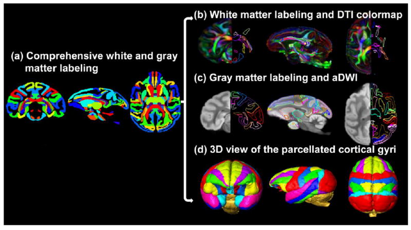
Comprehensive white and gray matter labels in the axial, parasagittal and coronal orientation. The labels of both white and gray matter are displayed in a typical axial, parasagittal and coronal plane in (a). In (b), the left and right sides of the coronal and axial planes show the population-averaged orientation-encoded colormaps and their corresponding WM tract labels, respectively. In (c), the left and right sides of the coronal and axial planes show the population-averaged aDWI images and their corresponding labels of cortical gyri and subcortical nuclei, respectively. The WM and gray matter labels are directly overlaid on a parasagittal plane of DTI colormap and aDWI image in (b) and (c), respectively. The anterior, lateral and superior view of the 3D reconstructed labeled cortical gyri are displayed in (d).