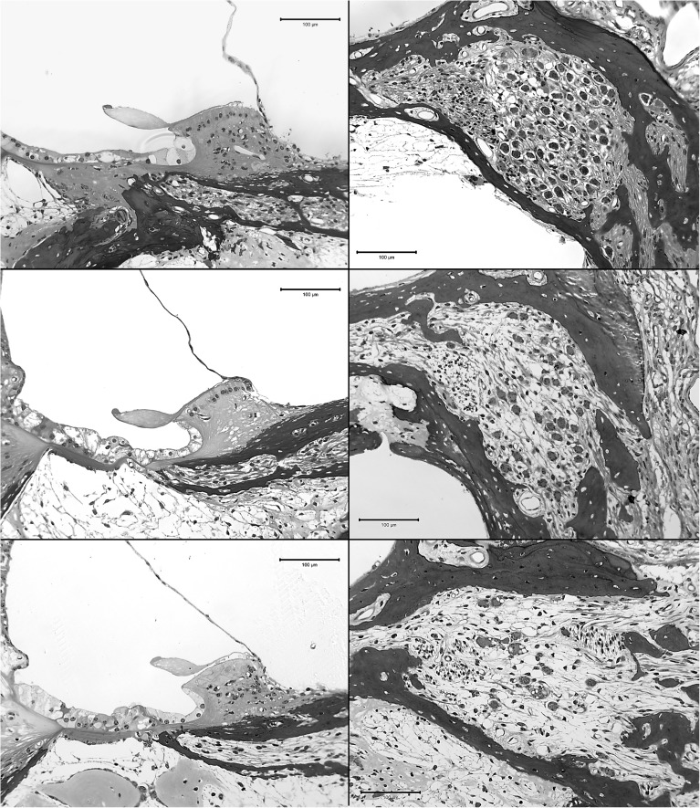
FIG. 2.
Examples of the basilar membrane area (left column) and SGN cell bodies in Rosenthal’s canal (right column) from profile A; examples for three cases are shown. Peri-midmodiolar sections were made at the location of the primary stimulating electrode in each case. Animal “g” (top row; survival time 183 dpi) had a neomycin injection into the scala tympani followed 30 min later by AAV.Ntf3 inoculation, 30 min before implantation. In this case, IHCs remained apical to profile A despite the neomycin injection. Animal “c” (middle row; survival time 377 dpi) received a similar treatment to animal “g” but no surviving IHCs were found in any of the profiles. A deafened-control animal (bottom row; survival time 176 dpi) had a neomycin injection followed by AAV.empty inoculation 30 min before implantation; no IHCs were found in any of the turns. SGN densities and other details for these three cases are plotted along with those for all other cases in the following figures.

