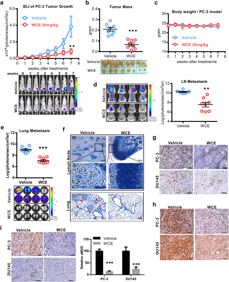
Figure 2.
Therapeutic effect of WCE on an orthotopic xenograft model of hormone-refractory PCa. (a) Longitudinal bioluminescence images (BLI) of PC-3-Luc2 prostate tumors in each group were measured weekly and are presented as a growth curve (n = 8). Bottom, the longitudinal images of BLI in representative mice. (b) The tumor weight following 7-week treatment is represented as a column scatter plot. Horizontal lines, mean (n = 8); Bottom, photograph of representative primary tumors taken at the endpoint. (c) Mouse body weight was monitored weekly. (d,e) Effect of WCE on tumor metastasis. BLI of the host lung (d) and lumbar lymph nodes (e) were examined at the endpoint. Metastasis was quantified by BLI as photon flux per second, and the results are represented as a column scatter plot (n = 8). (f) Immunohistochemical staining of metastases in lumbar lymph nodes and lungs for Ki-67 to indicate proliferating PCa cells. Arrows indicate cancer cells. (g) Anti-BrdU antibody was applied to determine proliferative cells in tumors. Scale bar, 100 μm. (h) IHC staining of HIF1α in PC-3 and DU145 tumor tissues. Scale bar, 50 μm. (i) The neovasculature of tumors was determined by CD31 staining in PC-3 and DU145 tumor slices and quantified as relative positive area (area microvessel density, aMVD) from four random fields in one tumor slice, n = 3 (right). Scale bar, 50 μm. All values are represented as means ± SEM. **P ≤ 0.01; ***P ≤ 0.001 (t-test, two-tail).

