Figure 4.
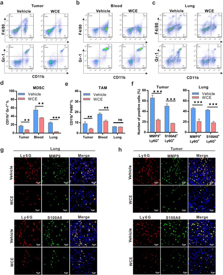
Effect of WCE on the mobilization and recruitment of myeloid cells in a nude mouse bearing PC-3 tumors. (a–c). The percentage of Gr-1+/CD11b+ myeloid cells and F4/80+/CD11b+ tumor-associated macrophages in primary tumors (a), peripheral blood (b) and lung (c) in tumor-bearing mice treated with WCE or vehicle as identified by FACS. (d,e) The percentage of MDSCs (d) and TAMs (d) in different tissues are shown as means ± SE (n = 3). (f) Fractions of Ly6G+/MMP9+ and Ly6G+/S100A8+ cells in primary PC-3 tumors (left) and in the lung (right) were calculated (n = 5). (g,h) The representative image of Ly6G+/ MMP9+ cells (g) and Ly6G+/S100A8+ cells (h) in primary PC-3 tumors and lungs were detected by immunofluorescent staining. Bar, 20 μm. All values are represented as means ± SEM. *P ≤ 0.05; **P ≤ 0.01; ***P ≤ 0.001 (t-test, two-tail).

