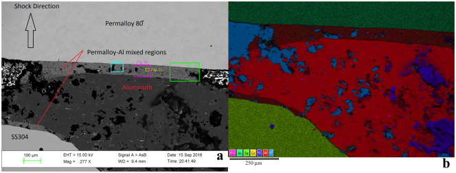
Figure 1.
General views of the polished surface of recovered specimen from shot S1235. First shock propagated from bottom of images towards top. (a) Backscattered electron image, showing the initially 1 mm thick Al2024 layer attenuated by flow associated with impact deformation to ~0.5 mm in this area near the left side-wall of the chamber. Regions of intermediate backscatter contrast mark a reaction zone up to ~0.1 mm wide with mean atomic number intermediate between the Al-rich and Ni-rich starting materials. (b) X-ray intensity map of the same general area, using the color scheme shown at lower left. Voids in the sample appear cyan due to Carbon in the epoxy fill. The brown regions mark the mixed layers.

