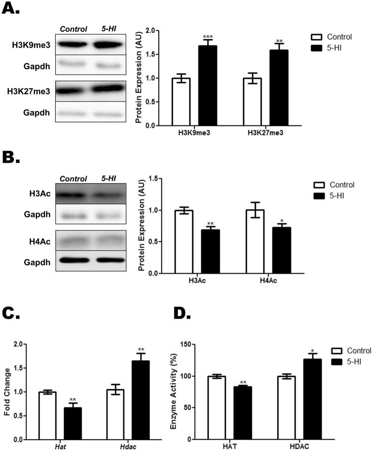
Figure 7.
Maternal IE exposure induces post-translational modifications in the thyroid of the adult male offspring. Thyroid lobes were obtained from the adult male offspring of control (C) or IE-exposed rat dams (5-HI). Thereafter, the methylation status of the lysines 9 and 27 of histone H3 (A) and acetylation status of the histones H3 and H4 (B) were evaluated through Western blotting analysis, using Gapdh as loading control (n = 8/group). Representative western blots are shown in the left panels. (C) Thyroid Hat and Hdac mRNA expression were analyzed by Real-Time PCR and normalized to Rpl19 mRNA content (n = 10/group). (D) Thyroid HAT activity was measured with a colorimetric assay kit, while HDAC activity was assessed with a fluorimetric assay kit. Results are expressed as means ± SEM as arbitrary units (AU) (A-B), fold change (C) or percentage (%) (D),. *P < 0.05, **P < 0.01; ***P < 0.001 vs. C (Unpaired two tailed Student’s t-test).

