Figure 1.
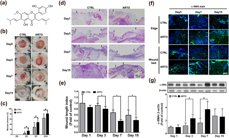
ARTO accelerates skin contraction and wound healing. (a) The structure of ARTO. (b,c) Representative photographs of wound healing in CTRL and ARTO-treated mice at various time points after wounding. The wound closure results were quantified on days 3, 7, and 15 after wounding. (d,e) H&E-stained sections on days 1, 3, 7, and 15 after wounding. The widths of the wound areas are marked by arrows and were quantified on days 1, 3, 7, and 15 after wounding. (f) Immunohistochemistry was performed to identify α-SMA in wounds on days 3, 7, and 15 after wounding. (g) The α-SMA levels were determined by western blot analysis. The data are shown as the means ± S.D. N = 6–18 wounds/group and *P < 0.05.

