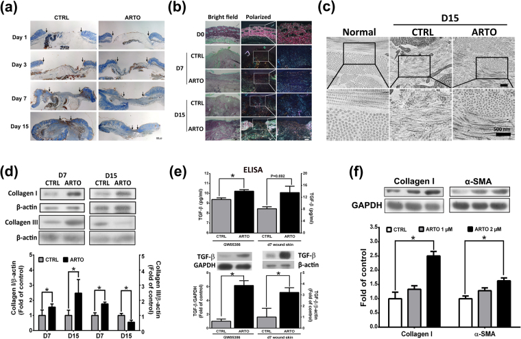
Figure 3.
ARTO enhances collagen deposition and maturation. The wounded sections stained with Mallory’s trichrome (a) and picrosirius red (b) showed that more newly formed and mature collagen fibers were observed in ARTO-treated wounds. The widths of the wound areas are marked by arrows. (c) TEM images of connective tissue on day 15 after wounding. Higher magnification images (lower panel) illustrate different collagen fibril diameters, amounts, and arrangements in skin wounds. (d) Western blot analysis of the skin showed collagen deposition (collagen type I and III) on days 7 and 15 after wounding. (e) TGF-β level was analyzed in GM05386 fibroblasts and skin by ELISA and western blot. (f) The collagen I and α-SMA levels were determined by western blot analysis. The data are shown as the means ± S.D. N = 3–6 wounds/group and *P < 0.05.

