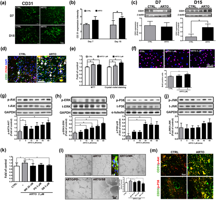
Figure 6.
ARTO enhances angiogenesis through the Akt or P38 signaling pathway. (a,b) Wounded sections were stained with CD31, and a quantitative analysis of CD31 level was conducted. (c) Western blot analysis of the skin showed that CD31 level was increased in the ARTO-treated wounds on day 15 after wounding. (d) Immunohistochemistry was performed to identify CD31 and PCNA (arrows) in wounds on day 15. (e,f) MTT, crystal violet staining, and BrdU incorporation assays were used to measure cell viability and proliferation. The levels of Akt (g), ERK (h), P38 (i), and JNK (j) were determined by western blot analysis. HUVECs were pre-treated with Akt or MAPK inhibitors for 1 h and then incubated with ARTO for 24 h. (k) Crystal violet staining was performed. (l) Tube formation and tube length were examined by a Matrigel assay. Confocal image of a tube stained for CD31 (green) and cell nuclei (DAPI, blue). The asterisk indicates the lumen of the tube. (m) There was co-localization between CD31 and phosphorylated Akt or P38 in the wounds on day 15. The data are shown as the means ± S.D. N = 3–6 wounds/group and *P < 0.05.

