Fig. 2.
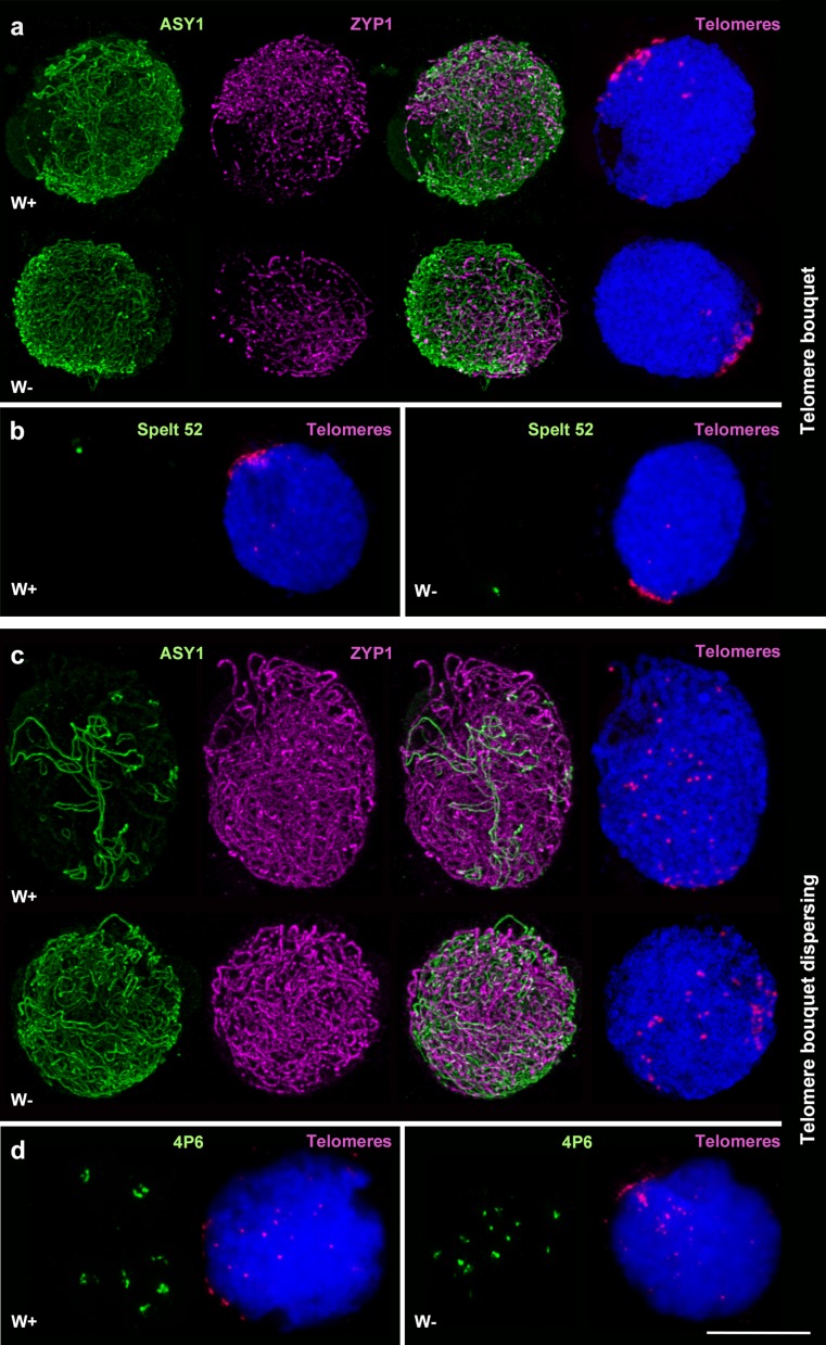
Progression of pairing and synapsis in wheat in the presence (W+) and absence (W−) of the Ph1 locus. a, c Immunolocalisation of meiotic proteins ASY1 (green) and ZYP1 (magenta) combined with telomeres (magenta) labelled by FISH in W+ and W− meiocytes. a During early telomere bouquet dispersal (group 2), levels of synapsis were similar in both W+ and W−. c When the telomere bouquet is dispersed but ASY1 and ZYP1 do not colocalise yet (group 3), levels of synapsis were higher in W+ revealing a delay in synapsis in W−. b FISH using Spelt 52 (green) and a telomere (magenta) probe during the telomere bouquet (group 1). Spelt 52 produce a single signal in the distal region of chromosome 4BS. Only one signal was observed in both W+ and W− during the telomere bouquet confirming the data shown in a. d FISH using 4P6 (green) and a telomere (magenta) probe during telomere bouquet dispersal (group 2). 4P6 labels seven interstitial sites on chromosomes of the D genome. In the figure, only 7 signals are observed in W+ during the telomere bouquet dispersal, while 12 signals are shown in W−. FISH results reveal a delay in pairing in the absence of Ph1, confirming the delayed synapsis shown in c. DAPI staining in blue. Scale bar represents 10 μm

