
Figure 6. mvsFIt inhibits VEGF165-driven HUVEC migration.
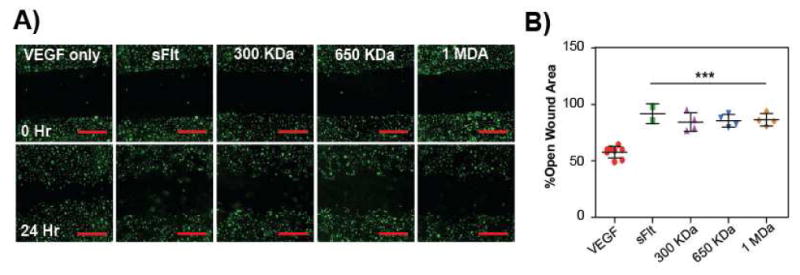
Representative images of inhibition of HUVEC migration with LCR mvsFIt bioconjugates of varying molecular weights. HUVECs were allowed to grow to confluence in 12-well plates prior to making a scratch and were treated with 20 ng/mL VEGF165 in the presence of 200 ng/mL mvsFIt. Cells were stained with CellTracker Green (Life Technologies) prior to seeding. Scale bar = 20 μm. B) Quantified HUVEC migration following treatment with LCR mvsFlt showing percent open wound area calculated by comparing open wound area at 24 hours to open wound area at time 0 (***p<0.001).