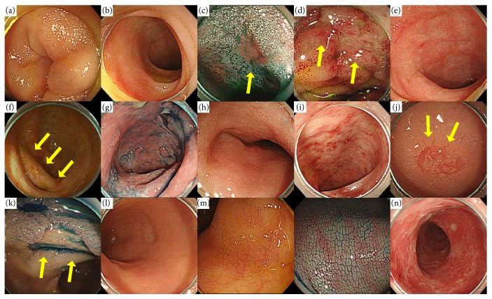
Figure 2.
Examples for endoscopic findings of lower GI: (a) edema, (b) redness, (c) erosion, (d) ulcer, (e) villous atrophy, (f) atrophy of the ileocecal valve, and (g) mucosal exfoliation in the terminal ileum and (h) edema, (i) redness, (j) erosion, (k) ulcer, (l) low vascular permeability, (m) tortoise shell-like mucosae, and (n) mucosal exfoliation in the colon.

