Figure 1.

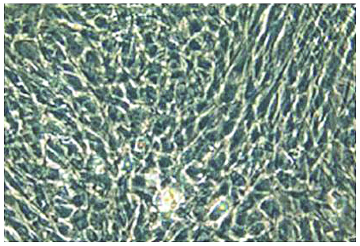
Morphological observation of second-generation chondrocytes after in vitro culture for 1 day. Inverted fluorescence phase-contrast microscopy showed that the cells were mostly polygonal with good growth status (×200).

Morphological observation of second-generation chondrocytes after in vitro culture for 1 day. Inverted fluorescence phase-contrast microscopy showed that the cells were mostly polygonal with good growth status (×200).