Figure 2.

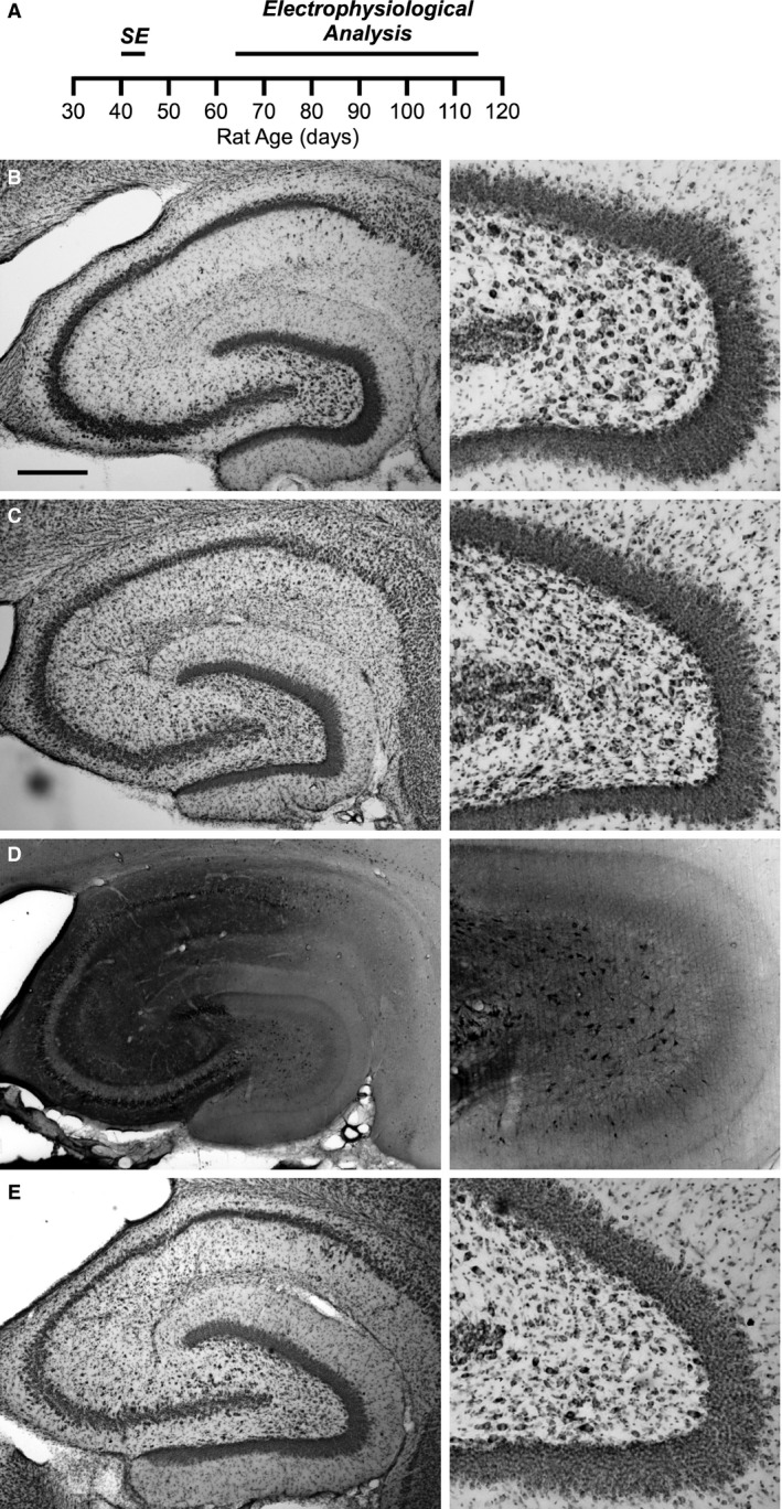
Pilocarpine treatment does not produce extensive neuronal cell death in the CA1 region of the hippocampus. (A) Timeline of the experimental protocol. Pilocarpine or saline is injected in to rats aged P40–P45 producing status epilepticus in the pilocarpine‐treated rats. Whole‐cell current clamp recordings from CA1 neurons were done 3–10 weeks later. (B–E) Nissl and Fluoro‐Jade B staining of the hippocampus (left) and expanded view of the hilus from the same slice (right). (B) Nissl stain 4 days following saline injection shows normal hippocampal structure. (C) Nissl stain 4 days following pilocarpine injection. There is no obvious reduction in Stratum pyramidale in CA1 and CA3 regions (left). However, characteristic damage resulting from status epilepticus can be observed in the hilar region (right). (D) Fluoro‐Jade B staining depicted in reverse grayscale of the hippocampus 4 days following pilocarpine injection from the same animal as in (C) Sporadic degenerating neurons (dark neurons) are visible throughout the hippocampus (left). Hilar neuron degeneration is also apparent (right). (E) Nissl stain 45 days following pilocarpine injection suggests that additional damage to the hippocampus did not occur after the acute response. Lack of degenerating neurons by Fluoro‐Jade B staining confirms this result (data not shown). Scale bar represents 0.5 mm.
