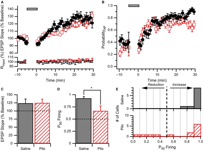
Figure 3.

WPP produces strong E‐S potentiation in neurons from saline but not pilocarpine‐treated animals. (A) Upper Plot: Average normalized EPSP slope time course for CA1 pyramidal cells from pilocarpine‐treated (open red squares) and saline‐treated (filled black circles) rats. Responses are normalized to the average baseline response before WPP (hatched bar). No difference in average EPSP slope time courses after WPP were observed between groups. Lower Plot: Average input resistance normalized to baseline for the same data sets. (B) Average AP firing probability (ProbabilityAP) time course for CA1 pyramidal cells from pilocarpine‐treated (open red squares) and saline‐treated (filled black circles) rats. No differences in average AP firing probability time courses were observed. (C) LTP induced by WPP is similar in neurons from saline and pilocarpine (Pilo) treated rats. EPSP slopes measured 30 min after WPP were normalized to their respective baseline values before WPP. (D) E‐S plasticity as measured by P50 Firing values in cells from pilocarpine‐treated rats was significantly lower than saline‐treated rats following WPP. * indicates significance P < 0.03. (E) E‐S plasticity is highly variable in CA1 cells from pilocarpine‐treated rats. Histogram showing that P50 Firing values analyzed from CA1 pyramidal cells from saline‐treated rats (solid) are clustered at high P50 Firing values, whereas those from pilocarpine‐treated rats (hatched) are more distributed.
