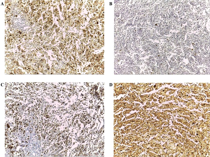
Figure 3.
Expression patterns of CT antigens in poorly differentiated carcinoma (magnification, ×100). (A) Immunohistochemical staining with mAb Multi MAGE-A with (++++) immunoreactivity. (B) Immunohistochemical staining with mAb CT7-33 with focal immunoreactivity. (C) Immunohistochemical staining with mAb #26 with (++++) immunoreactivity. (D) Immunohistochemical staining with mAb E978 with (++++) immunoreactivity. CT antigen, cancer/testis antigen; mAb, monoclonal antibody; MAGE-A, melanoma-associated antigen A; MAGE-C1/CT7, melanoma-associated antigen C1.

