Fig. 1.

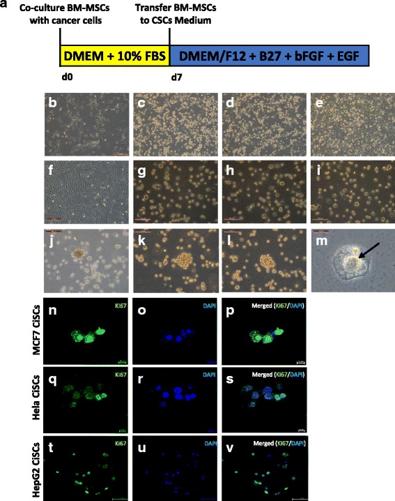
Generation of CiSCs from adult BM-MSCs. a Schematic illustrating the time schedule and derivation of CiSCs from BM-MSCs. b Morphology of BM-MSCs at day 0 cultured in standard conditions. c, d BM-MSCs after 2 days in coculture with (c) MCF7, (d) Hela, and (e) HepG2 cells. f Morphology of BM-MSCs at day 5 cultured in standard conditions without cancer coculture. g–i BM-MSCs after 5 days in coculture with (g) MCF7, (h) Hela, and (i) HepG2 cells showing generation of spheroid-like cells. j–l CiSCs growing in colonies in suspension. m Central cavity formation (arrow) becomes evident in CiSCs after several weeks in culture. n–v Confocal immunofluorescence images for Ki-67 of (n–p) MCF7, (q–s) Hela, and (t–v) HepG2 CiSCs. Nuclei stained with DAPI (blue). BM-MSC bone marrow mesenchymal stem cell, CiSC cancer-induced stem cell, CSC cancer stem cell, bFGF basic fibroblast growth factor, DMEM Dulbecco’s modified Eagle’s medium, EGF epidermal growth factor, FBS fetal bovine serum
