Fig. 2.

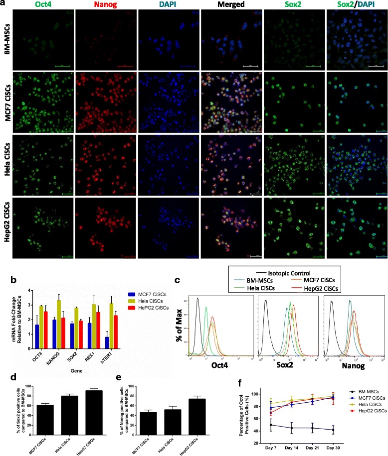
CiSCs express human ESC-specific markers. a Confocal immunofluorescence images for Oct4 (green), Nanog (red), and Sox2 (green) of control BM-MSCs and MCF7, Hela, and HepG2 CiSCs. Nuclei stained with DAPI (blue). Scale bars = 60 μM. b Expression levels of mRNAs encoding OCT4, NANOG, SOX2, REX1, and hTERT in MCF7, Hela, and HepG2 CiSCs relative to parental BM-MSCs determined by real-time qRT-PCR. Data reported on a log-10 scale as mean ± SD. c Flow cytometry overlay histogram analysis of Oct4, Sox2, and Nanog in BM-MSCs and MCF7, Hela, and HepG2 CiSCs. For comparison, isotype control (black) was used to define the positive and negative populations for each marker. d Oct-4 protein expression levels increase with the number of days (7–30) when cultured in the presence of B27, EGF, and bFGF, as determined by intracellular flow cytometry staining, indicating the self-renewal capacity of CiSCs. e, f Quantification of the percentage of (e) Sox2-positive and (f) Nanog-positive cells compared to parental BM-MSCs by intracellular flow cytometry staining. Proportions of positive cells measured by subtracting control parental BM-MSC staining from test histograms using super-enhanced Dmax (SED) normalized subtraction using FlowJo v. 10.2 software. Data presented as mean ± SD. BM-MSC bone marrow mesenchymal stem cell, CiSC cancer-induced stem cell
