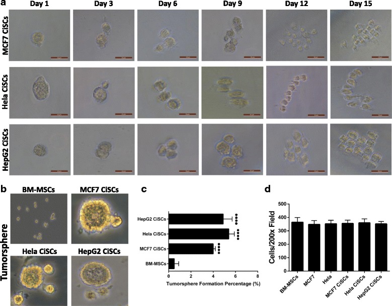
Fig. 3.

Single-cell colony formation, tumorsphere formation, and invasiveness of CiSCs. a Representative phase-contrast images of single CiSCs plated at a clonal density by limited dilution assays showing the colony-forming efficiency of a single CiSC. b Phase-contrast images of tumorspheres formed from BM-MSCs and MCF7, Hela, and HepG2 CiSCs. c Quantification of tumorsphere-forming ability of BM-MSCs and MCF7, Hela, and HepG2 CiSCs showing CiSCs to have significantly higher tumorsphere formation percentage (P < 0.05). Data presented as mean ± SD. d Quantification of invading cells toward lower chamber of the insert (average of 10 picture fields at 200× total magnification for each cell type). Data presented as mean ± SD. BM-MSC bone marrow mesenchymal stem cell, CiSC cancer-induced stem cell
