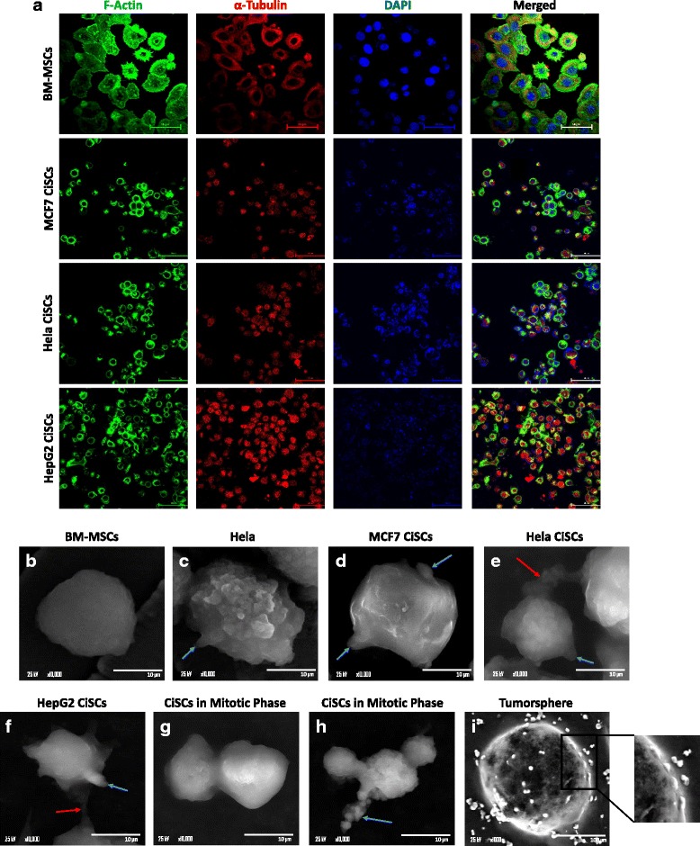
Fig. 8.

Cytoskeleton organization and surface ultrastructural characterization of CiSCs. a Confocal immunofluorescence staining of the actin cytoskeleton using phalloidin (green) and α-tubulin (red) showing localization of actin around the cell periphery while the α-tubulin network was distributed throughout the cell. Nuclei stained with DAPI (blue). Scale bars = 60 μM. b–i SEM revealed parental BM-MSCs to have a smooth and uniform surface (b) while (c) Hela cancer cells and (d) MCF7, (e) Hela, and (f) HepG2 CiSCs had an irregular surface and many microvilli and protrusions in the form of tumor-like buds (blue arrows). Adjacent cells interconnected by active pseudopodia (red arrows). g, h Mitotic cell division phase of CiSCs showing apophysis. (i) SEM of a tumorsphere and magnification showing tumor-like buds on the surface of the tumorsphere. BM-MSC bone marrow mesenchymal stem cell, CiSC cancer-induced stem cell
