
Fig. 9.
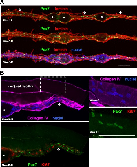
Pax7 staining. Confocal images of regenerating human skeletal muscle fibres. a A single muscle fibre is displayed, stained for laminin (red), Pax7 (green) and nuclei (blue), as maximum intensity projections of several slices to show that Pax7+ cells are located in both the regenerating zones (arrows) and at the periphery of the necrotic zones (asterisk). b Three fibres are visible, and the lowest fibre is undergoing regeneration. In the regenerating zone, it is clear that none of the Pax7+ cells is proliferating (Ki67+). The area indicated by the dashed line box is shown at higher magnification, where a cluster of Pax7+ cells was observed on the surface of the uninjured fibre. Scale bars, 100 μm