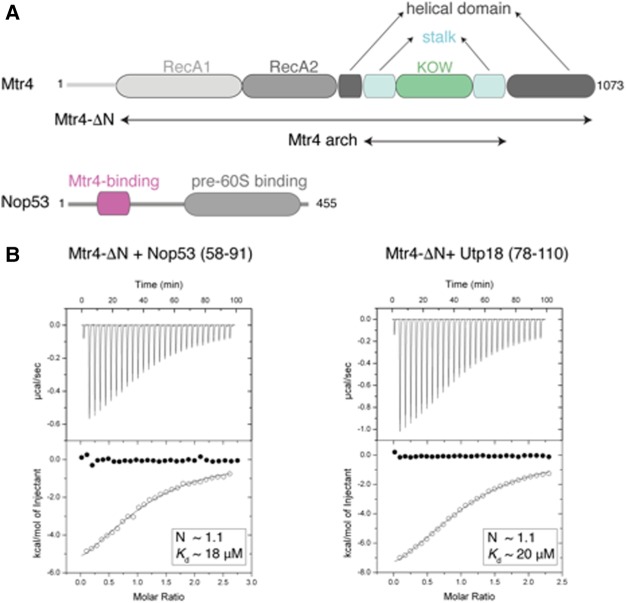
FIGURE 1.
Biophysical characterization of Mtr4 binding to AIM-containing proteins. (A) Schematic representation of the domain structure of yeast Mtr4 and Nop53. Predicted unstructured regions are represented as gray lines. The Nop53 fragment identified by proteolysis contains the arch-interacting motif (AIM) identified by Thoms et al. (2015). (B) ITC experiments of Mtr4-ΔN with Nop53prot and Utp18prot. The open circles show the titration of Nop53prot/Utp18prot into the Mtr4-ΔN containing cell. The filled circles show the control where Nop53prot/Utp18prot were titrated into buffer. In each inset is the number of calculated binding sites (N), and dissociation constants (Kd) are shown.

