Fig. 3.

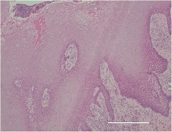
Pathological microscopic examination reveals thickened squamous epithelia with slight nuclear atypism and disorders of the epithelial rete pegs accompanied by moderate grade inflammatory cell infiltration (HE staining, bar: 400 μm)

Pathological microscopic examination reveals thickened squamous epithelia with slight nuclear atypism and disorders of the epithelial rete pegs accompanied by moderate grade inflammatory cell infiltration (HE staining, bar: 400 μm)