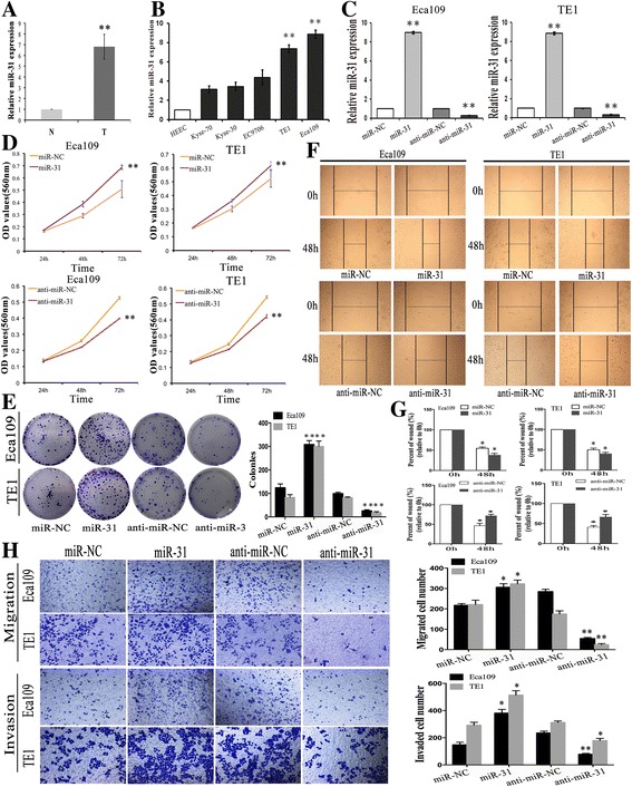
Fig. 1.

Expression of miR-31 in ESCC cell lines and tissue samples and the in vitro effects of miR-31 on cell proliferation, migration and invasion in ESCC cells. a The relative expression level of miR-31 in 20 specimens of ESCC (T) and the adjacent nontumor tissues (N) was determined by qRT-PCR. b QRT-PCR analysis miR-31 expression in five human ESCC cell lines and the normal esophageal epithelial cell line (HEEC). c QRT-PCR analysis of the relative expression of miR-31 in each group of ESCC cells transfected with miR-31 mimics and inhibitor. d-e MTT and colony formation assays in ESCC cells overexpressing or underexpressing miR-31. f-g Wound scratch healing assay of ESCC cell showed that change of miR-31 effectively affected cell motility. Photographs were taken immediately (0 h) and at 48 h after wounding, quantification of wound closure was done. h Migration assay and Invasion assay revealed that the upexpression or downexpression of miR-31 promoted or inhibited the invasion ability of ESCC cells. Results are expressed as the mean ± SD of three independent experiments. *: P < 0.05; **: P < 0.01
