Fig. 3.

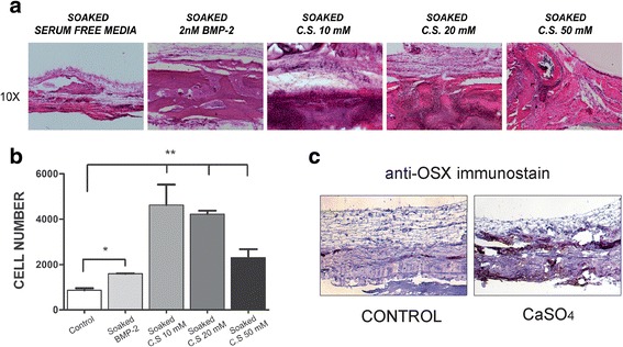
CaSO4 increases bone regeneration in vivo by recruiting the host’s cells into the bone defect. a HE staining shows host’s cells recruited into the cell-free implanted scaffold. Representative images of the control group (soaked in serum-free media), 2 nM BMP-2, CaSO4 (C.S.) 10 mM, C.S. 20 mM, and C.S. 50 mM taken from the center of the defect. 10×, scale bar = 400 μm. b Recruited cells into the different scaffolds quantified as described in Methods. c Osteoprogenitor cells expressing Osterix (OSX) identified in a representative implanted control and calcium-containing scaffold by immunohistochemistry. Differences considered significant at *p < 0.05, **p < 0.01. BMP bone morphogenetic protein
