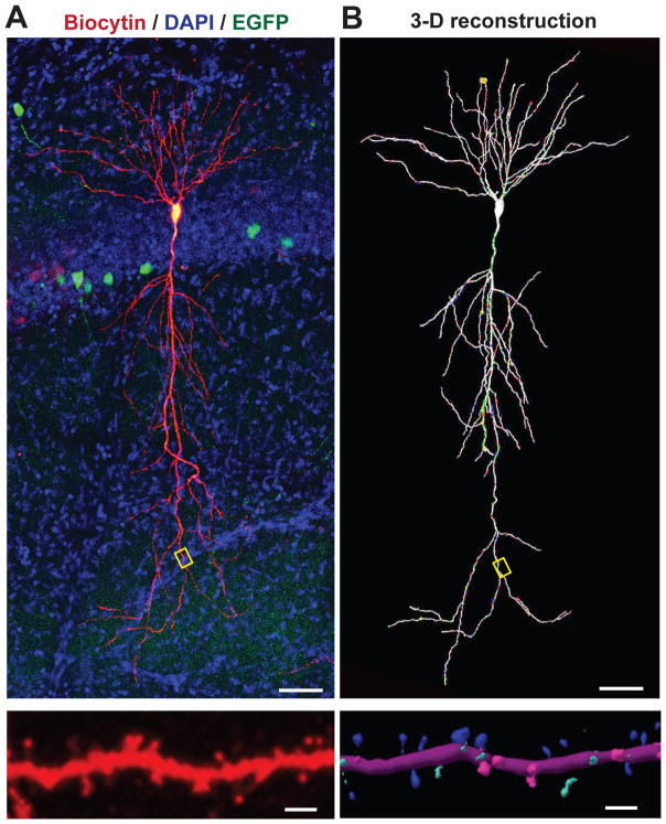
Figure 1. Illustration of neuronal shape and spines: An exemplary pyramidal neuron from the CA1-region of the hippocampus with spine-studded dendritic extensions.
A. Top, a CA1-region pyramidal neuron was patched in hippocampal slices from a mouse that had been sparsely infected with a lentivirus encoding EGFP, and was filled with biocytin via the patch pipette (a section stained for biocytin (red) and DAPI to label nuclei (blue). Bottom, expansion of the dendritic field boxed in the neuronal overview image above to illustrate the dense decoration of dendrites with spines.
B. 3D-reconstruction of the pyramidal neuron and its dendritic segment shown in A. In the bottom panel, spine shapes were categorized and color–coded (blue, mushroom; green, thin; pink, stubby). Images courtesy of Dr. Richard Sando.

