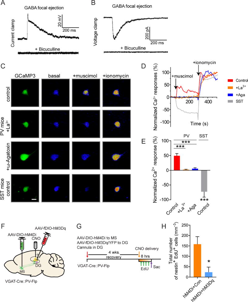
Figure 4. GABA induces depolarization and Ca2+ influx in PV interneurons in the adult dentate gyrus.
(A–B) Gramicidin-perforated patch-clamp recordings of dentate PV neurons in response to GABA focal ejection (5 µM) with or without bicuculline (50 µM) under the current-clamp (A) and voltage-clamp mode (B) (n=6). (C–E) Ca2+ imaging analysis of neuronal responses to GABAAR agonist muscimol (10 µM) and Ca2+ ionophore ionomycin (10 µM). Adult PV or SST neurons were labeled with AAV expressing GCaMP3 in PV-Cre or SST-Cre mice. (C) Confocal images of Ca2+ responses without or with pre-treatment of La3+ (50 µM) or ω-Agatoxin TK (100 nM). Scale bar: 20 µm. (D) Calcium time course analysis. (E) Summary of the mean peak Ca2+ responses. Values are normalized to the mean fluorescence intensity at baseline (0%) and after ionomycin (100%). (n = 7 cells from >3 mice for each group). (F) Viral injection and DG cannulation scheme in VGAT-Cre::PV-Flp mice. (G) Experimental paradigm for in vivo CNO infusion. (H) Density of activated rNSC (nestin+EdU+). (n=5 for hM4Di+Con, and n=4 for hM4Di+hM3Dq). *p<0.05, ***p < 0.001 by Student’s t-test. Values represent mean ± S.E.M. See also Figure S4.

