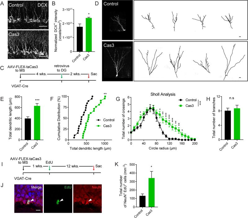
Figure 7. Chronic ablation of medial septum GABA neurons leads to impaired hippocampal neurogenesis.
(A) Confocal images showing DCX cells in the DG in control and caspase conditions at 6 wpi. Scale bar: 50 µm. (B) Density of normalized fluorescence intensity of DCX immature neurons (n=4 for control and caspase). (C) Experimental scheme of chronic ablation of MS GABAergic neurons and retroviral birth-dating of adult born neurons in VGAT-Cre mice. (D) Representative dendritic trees of GFP+ newborn neurons. Scale bar: 10 µm. (E) Total dendritic length (n=20 cells from 4 mice for both control and caspase). (F) Cumulative distribution of total dendritic length (same dataset from E). **p<0.01 by Kolmogorov-Smirnov test. (G) Sholl analysis of GFP+ newborn neurons at 14dpi (same dataset from E). (H) Total number of dendritic branches (same dataset from E). (I) Experimental scheme of chronic ablation of MS GABAergic neurons in VGAT-Cre mice. (J) Confocal images of mature neurons (NeuN+EdU+) following 13 weeks of MS GABA neuron ablation. Scale bar: 10 µm. (K) Density of mature neurons (NeuN+EdU+) (n=4 for control and caspase). *p<0.05, **p<0.01, ***p<0.001 by Student’s t-test. Values represent mean ± S.E.M. See also Figure S7.

