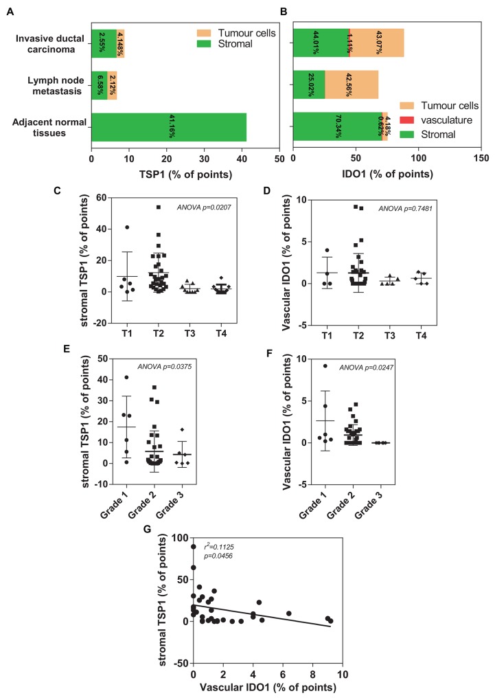
Figure 4. Histomorphometric analysis of TSP1 and IDO1 in a TMA of human invasive ductal carcinoma patients.
(A) Plot of percentage of immunostaining TSP1 covering different areas on tissue microarray, including adjacent normal tissues, invasive ductal carcinoma and lymph node metastasis. (B) Plot of percentage of immunostaining IDO1 covering different areas on tissue microarray, including adjacent normal tissues, invasive ductal carcinoma and lymph node metastasis. (C) A significantly association between decreased histomorphometric scores of the stromal TSP1 and high TNM status (p=0.0207). (D) A trend toward reduced histomorphometric scores of the vascular IDO1 was observed in patients with IDC who had an overall poor outlook (T3 and T4), compared with those patients with a relative good prognosis (T1 and T2). (E) Decreased histomorphometric scores of the stromal TSP1 was significantly correlated with poor differentiated grade tumours (p=0.0375). (F) Histomorphometric scores of the vascular IDO1 significantly decreased in IDC tissues with poor differentiated grade with the observation that the lower vascular IDO1 were in high TNM status (p=0.0247). (G) Plot of the vascular IDO1 (histomorphometric scores as % of points) on the horizontal axis versus the stromal TSP1 (histomorphometric scores as % of points) on the vertical axis. The trend in the points is given by the line with as statistical significance (R2=0.1125, p=0.0456).

