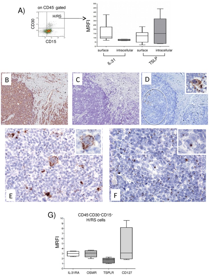
Figure 1. Expression of IL-31, TSLP and their receptors in H/RS cells.

(A) Left panel. A representative gating strategy for H/RS cells identified as CD45, CD30+, CD15+ cells. Right panel. IL-31/TSLP expression was tested by flow cytometry at surface and intracellular levels. Results are expressed in box plot as median MRFI, first and third quartiles, maximum and minimum values, from 10 different HL lymph node cell suspensions. (B-F) In situ hybridization for Ubiquitin (B), dapB (C), CD30 (D), IL-31 (E) and TSLP (F) mRNA in cHL using the RNAscope technology (B, C, D) original magnification x100; E, F x200; insets x400). Ubiquitin mRNA was diffusely expressed (brown dots), whereas the bacterial dapB was completely negative. The CD30 probe hybridized with a proportion of the cells with H/RS morphology (circle and inset). Both IL-31 and TSLP mRNA were detected in the cytoplasm of H/RS cells (inset) and in some of the immune reactive cells present in the background. (G) IL-31RA/OSMR and TSLPR/CD127 chain receptor expression was analyzed by flow cytometry. Results are expressed in box plot as median MRFI, first and third quartiles, maximum and minimum values, from 7 different HL lymph node cell suspensions.
