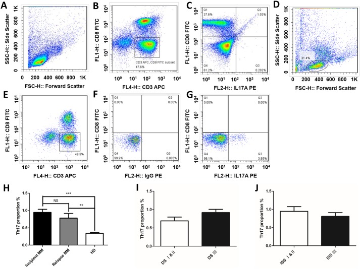
Figure 2. Th17 cells is correlated with the MM clinical stages.
The ratios of Th17 cells in peripheral blood (CD3+ CD8- IL-17A+) were detected by flow cytometric analysis in MM patients and healthy donors. The proportions of Th17 cells of normal people (A) the gate for lymphocytes was set by the FSC and SSC. (B) CD3+CD8- lymphocytes were selected by CD3 and CD8 door. (C) The ratio of CD3+ CD8- IL-17A+ in CD3+ CD8- lymphocytes. The proportions of Th17 cells of MM patient (D) the gate for lymphocytes was set by the FSC and SSC. (E) CD3+CD8- lymphocytes were selected by CD3 and CD8 door. (F) The same type of control chart for IL-17A. (G) The ratio of CD3+ CD8- IL-17A+ in CD3+ CD8- lymphocytes. (H) The proportions of Th17 cells in CD3+CD8- lymphocytes were detected by flow cytometric analysis in MM patients, the results showed that Th17 cells in the incipient and relapsed patients were significantly higher than normal (**p < 0.01, ***p < 0.001), Th17 cells in the incipient patients were higher than that in patients with recurrence, but there was no significant difference between them (p > 0.05). The percentages of Th17 cells in DS stage and ISS stage patients (I) DS stage; (J) ISS stage.

