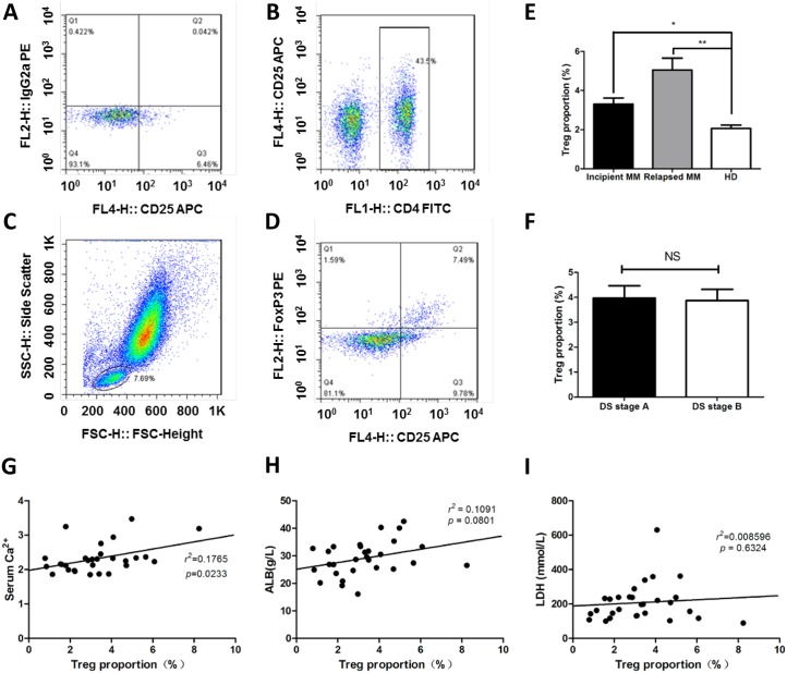
Figure 4. The proportions of Treg cells is increased in MM patients.
The ratios of Treg cells in peripheral blood (CD4+CD25+FoxP3+) were detected by flow cytometry in MM patients. (A) The gate for lymphocytes were set by the FSC and SSC. (B) CD4+ lymphocytes were selected by CD4 and CD25 door. (C & D) the ratio of CD4+CD25+FoxP3+ Treg cells in CD4+ lymphocytes. (E) The proportions of Treg cells in CD4+ lymphocytes were detected by flow cytometry, the results showed that Treg cells in the incipient and relapse patients were significantly higher than normal (3.23 ± 1.69% vs. 2.16 ± 0.71%, 5.06 ± 2.41% vs. 2.16 ± 0.71%, p < 0.01), Treg cells in the relapsed patients were higher than that in incipient patients (5.06 ± 2.41% vs. 3.23 ± 1.69%, p < 0.01). (F) The percentage of Th17 cells in DS stage. (G-I) Correlations of clinical parameters with Treg cells in incipient MM.

